The Ernst Ruska-Centre for Microscopy and Spectroscopy with Electrons (ER-C) has, for almost 20 years, dedicated itself to the task of imaging and analyzing materials down to the atomic and molecular level. It is now entering a new era.
The project name ER-C 2.0 suggests an update as would be usual with software. “But to describe our project as a simple improvement would be an understatement,” says Prof Carsten Sachse, one of the three ER-C directors. “It’s about more than that. As a national research infrastructure (see “Of major importance” at the end of the article), we are entering a new dimension – with new devices, new applications and new opportunities for science and industry.”
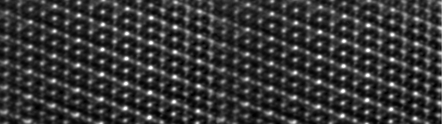
Five globally unique electron microscopes (see “Microscope profiles”) are designed to elicit information from materials and biological samples that is more detailed than any previously available. For example, they look for the position and chemical state of individual atoms or changes in the structure of substances that take place within a few femtoseconds – that is, one quadrillionth of a second. Detailed insights like these enable scientists to develop innovative materials – such as for energy transition – or new medicines more quickly.
Microscope profiles
TOMO
BIO
SPECTRO
OPERANDO
FEMTO
Atom probe tomography and high-resolution electron microscopy are combined in one device for the first time. With this, it should become possible to precisely determine the type and position of millions of atoms in a volume of several thousand cubic nanometres with a single measurement.
The first microscope cooled with a helium cryostat, which is equipped with lens correctors to make atoms in biological samples visible at -250°C.
The first ultra-low temperature in-situ microscope with ultra-high energy resolution. It will be used to analyze the electronic structure and vibrations of radiation-sensitive samples and polymers and to investigate atomic or molecular processes.
Ultra-high vacuum microscope to study materials while they are in use – for example during the operation of a battery.
Microscope with femtosecond resolution to analyze dynamic and ultrafast processes in the nanometre range.
Electron microscopes make tiny details visible
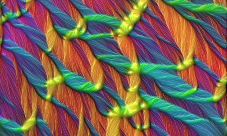
The colors show the different directions of the magnetic field in a 20 nanometer thin layer of polycrystalline cobalt.
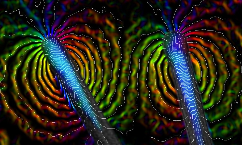
Nanotubes: The colors stand for the direction and intensity of the field, the contours for the magnetic field lines.
Magnetic field lines around the end of a magnetized iron wire
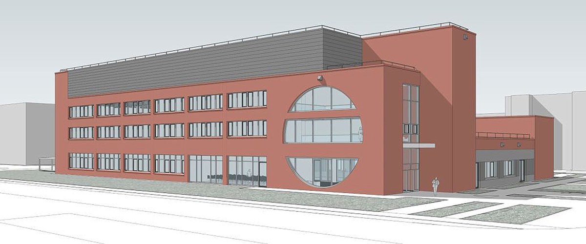
The sensitive electron microscopes need a perfect environment for maximum performance: free from vibration, shielded from electromagnetic radiation and with a constant temperature. Due to be fully operational in autumn 2024, a €23 million research building offers exactly that. Among other things, its foundation is a huge concrete block up to 1.5 metres thick, which ensures that vibrations from passing lorries, for example, do not distort the measurement results.
Everything under one roof
However, the new building is not only designed for the microscopes. “Until now, the scientists at ER-C have been spread across several buildings. The fact that we will be under one roof in the future and have more space for interdisciplinary exchange, for example, is of great value,” says the director, Prof. Joachim Mayer. The head of Materials Science and Technology is convinced that innovations often result from building bridges between disciplines.
There are three that benefit from one another in ER-C: the two traditional disciplines of materials science and solid state physics, plus the life sciences, which were added around five years ago. “With highly sensitive methods, electron microscopy can also be used for biological and medical questions,” says Sachse, head of Structural Biology. Electron microscopes can today be used to determine the structure of the body’s own proteins and see, for example, how drugs bind to them.
Of major importance
Large-scale equipment and instruments, but also data collections, computer networks and meeting centres, for example, are classified as research infrastructures. Large-scale devices such as electron microscopes in particular enable promising basic research and technological advances, but are generally expensive, complex and time-consuming to maintain. The Federal Ministry of Education and Research is funding selected infrastructures that are of major importance for Germany as a centre of science with a national roadmap – a kind of schedule for the long-term orientation of cutting-edge research. The infrastructures funded in this programme each receive over €50 million.
Access to the latest devices
In the course of its almost 20-year history, ER-C has repeatedly worked with companies and academic partners to advance microscopes and methods. “The companies supply us with their latest devices. Our scientists test them, develop new software and then give feedback on what they noticed,” explains the director, Prof. Rafal Dunin-Borkowski, head of Physics of Nanoscale Systems. “Based on this feedback, the companies provided us with improved versions of the devices, so we had access to new technologies before anyone else did – a perfect example of knowledge and technology transfer that benefits research and application.”
The ER-C directors can now expand their collaboration with the companies even further – by developing new cooling technologies, for example, to investigate quantum materials: “The funding associated with the inclusion of ER-C 2.0 in the national roadmap for research infrastructures opens up much better opportunities for us on this,” says Dunin-Borkowski. “The new era can begin.”
Contributions to structural change
ER-C 2.0 provides a valuable incentive for companies to locate in the Rhineland region. With its high-performance microscopes in one place, ER-C 2.0 offers, for example, the IT and energy technology industries unique opportunities to analyze and advance materials for energy storage or quantum processors. Pharmaceutical and medical companies settling down in the region can also receive support. This is not just a matter of equipment and expertise, but also of a highly qualified workforce. The Centre also specifically promotes the willingness of its scientists to spin off companies. Microscope manufacturers and their suppliers also benefit from it, for example through contact with the numerous users of the devices. Jülich, RWTH Aachen University and Heinrich Heine University Düsseldorf are involved in setting up the national competence centre for high-resolution electron microscopy and are cooperating with various partners.
Model of the new research building of "Ernst Ruska-Centre 2.0"Copyright: — pbr Planungsbüro Rohling AG
Text: Frank Frick | images: Juri Barthel/Forschungszentrum Jülich; Forschungszentrum Jülich/Ralf-Uwe Limbach; A. Husmann, M. McCartney, C. Boothroyd, R. Dunin-Borkowski; E. Simpson, Y. Hayashi, T. Kasama, R. Dunin-Borkowski; J. Thong, K. Harada, A. Tonomura, T. Akashi, T. Matsuda, Y. Togawa, C. Boothroyd, R. Dunin-Borkowski