The long summer of AI
The artificial intelligence boom has only just started. With its unique computer infrastructure and technical expertise, Jülich can make an important contribution.

Amino acid PET is an important tool in diagnosing brain tumours. However, determining the exact size of a tumour takes time and is not routine. An AI is set to change that. It can analyze PET images as well as experienced doctors – only much faster.
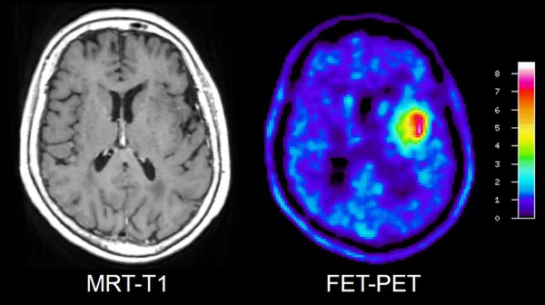
For the diagnosis and treatment of a brain tumour, it is important to determine the exact size and volume in order to be able to check whether the tumour is responding to treatment or whether it is continuing to grow. Clinics often use magnetic resonance imaging (MRI) for this, which specifically records structural changes in the tissue. However, these do not necessarily reflect the actual extent of the tumour. Another imaging procedure records the altered metabolism of the cancer cells and often provides results that differ from MRI: amino acid positron emission tomography (PET).
On the downside, determining the tumour volume using PET scans is very time-consuming. This is why the method is rarely used routinely. In the future, this is set to change – thanks to artificial intelligence (AI).
How PET works
PET uses radioactively labelled biomolecules to make metabolic processes visible. Amino acids have proven particularly useful for the imaging of brain tumours. The rapidly growing cancer cells take up the amino acids much faster than the healthy brain cells. Based on the enriched amino acids, the PET images can be used to determine the location and size of the metabolically active tumour.
Researchers from Jülich, Heidelberg and Cologne have developed a deep learning algorithm called JuST_BrainPET* that automatically recognizes brain tumours on PET scans and determines their volume. The team used 699 PET scans from 555 brain tumour patients for this purpose. The AI results agree very well with the values that experts determine from PET scans. Plus: the AI only needs a few minutes to do this.

The research team also had the algorithm assess the chances of success of treating patients with gliomas, which are the most common malignant brain tumours. “The clinical assessment of the AI as regards the response to therapy and prognosis was just as good as that of a specialist – and it took only a fraction of the time,” says principal investigator Priv.-Doz. Philipp Lohmann from the Institute of Neurosciences and Medicine (INM-4). “Our no-cost, freely available AI tool can’t replace doctors, but it can support them. We hope that it will encourage doctors to use amino acid PET more frequently with brain tumour patients – especially if they have little experience with the method.”
Text: Janosch Deeg | illustration (created with the help of artificial intelligence): SeitenPlan with Stable Diffusion and Adobe Firefly | image: Michael Wodak/MFK
* JUST_BrainPET: Juelich Segmentation Tool for Brain Tumor PET