ELECTRON MICROSCOPE

Electron microscopes (EM) allow tiny structures to be made visible like a gigantic magnifying glass - right down to individual atoms! They are now standard in biology and materials research, for example.
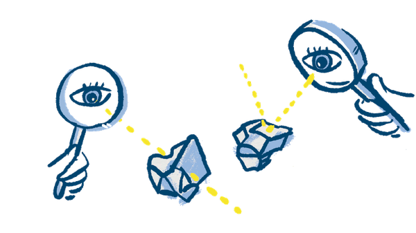
High Resolution - Huh?

Electron microscopes do not use a beam of light like conventional microscopes, but a beam of electrons.

Electrons have a wavelength up to a million times smaller than light - and the smaller the wavelength, the higher the resolution.

This makes it possible to recognize the atomic structure of a substance - that is, to decode structures that are only a few nanometers in size.
1931
Ernst Ruska and Max Knoll present the first electron microscop.
Ruska will bei awarded the Nobel Prize for this in 1986.
Illustrations: Diana Köhne


Last Modified: 16.02.2024